On the plate is a cross-section of the eye. The crystalline lens thickens, then relaxes. A beacon beam leaves the macula, and the wavefront exits the eye towards the sensor. The sensor is the letter D itself: inside it you can see the grid of microlenses, the astigmatism map turns, and the scan line runs from top to bottom. The mark on the scale moves from zero to −2.50 D.
Dioptre
In half a minute a laser removes a layer thinner than a hair from the cornea, and the eye starts to see without glasses. The surgeon works out how much to remove in dioptres. With a 6 mm optical zone, one dioptre of myopia is about 12 µm of tissue. The whole eye refracts light with a power of about 60 D, and an error of one sixtieth is already enough to blur the letters on the chart.
01 · Definition
Lens power is the reciprocal of focal length
A lens with a focal length of one metre has a power of 1 D. A focal length of half a metre gives 2 D, ten centimetres gives 10 D. A diverging lens gets a minus sign. Glasses for myopia have minus lenses, glasses for hyperopia have plus lenses.
The beauty of dioptres is that they add up. Light from a point 0.5 m away reaches the eye with a vergence of −2 D: that is what opticians call the curvature of the wavefront. The cornea adds its 43 D, the crystalline lens about 20 D more. If the sum brings the front to a focus exactly on the retina, the image is sharp. If not, the difference in dioptres is the prescription.
Inside the eye, light travels in a medium with an index of 1.336, so there vergence is divided not by the distance but by the distance divided by n. The cornea and the optical centre of the lens are almost 6 mm apart, and this gap also changes the sum. The lab below works through all of this step by step.
Measure the eye, correct it and see the world as it does
On the left is a cross-section of the eye at true size: cornea, crystalline lens, length from cornea to retina. Blue rays come from the object, violet ones are the two principal meridians in astigmatism, and the coral dot is where they meet. Top right is the patient's view: the picture is blurred by the eye's point spread function. Bottom right is the screen of the selected instrument. Below the scene is the visual acuity scale, logMAR.
02 · Conversion
Dioptres, focal length and old lens numbers
| {n} | {v} | {note} |
03 · Orders of magnitude
04 · Measuring instruments
05 · Writing rules
06 · Neighbouring units
07 · Historical section
The Jesuit Christoph Scheiner pricked two holes in a card and looked at a needle through them. If the eye is in focus, there is one needle. If not, there are two, and the way they split shows which way the error goes. Almost all autorefractors still work on this principle: the instrument finds the power at which the two beams meet at one point on the retina.
Catalogue of quantities
the whole index →The seven SI base units
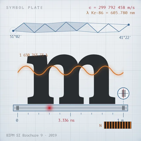
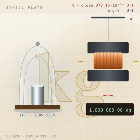
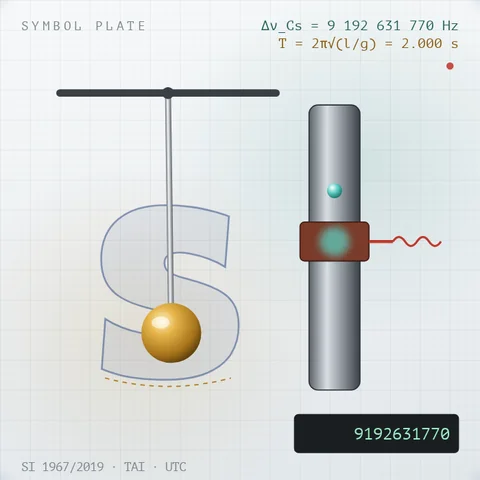
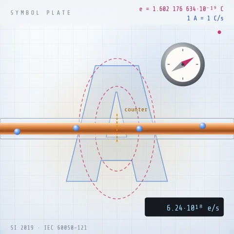
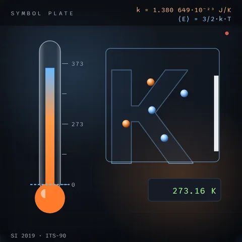
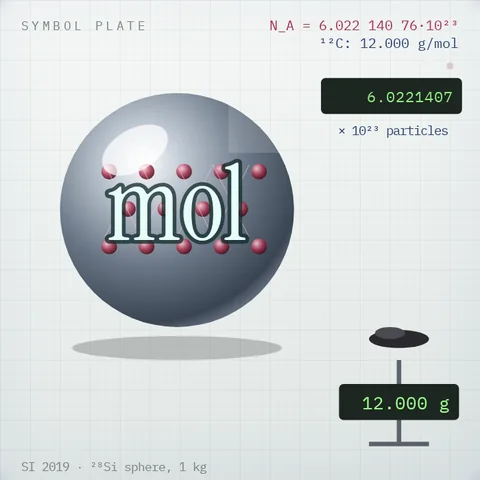
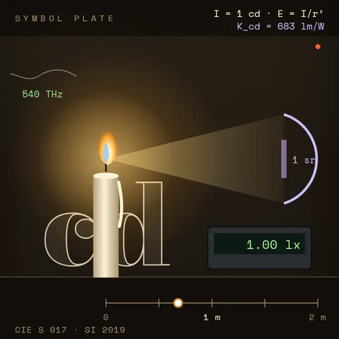
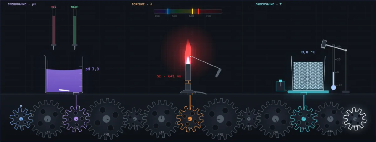
A drop of acid changes the colour of the juice, salt colours the flame, water freezes at a tap. Below them are all Simetrium sheets, by discipline and alphabetically.
Open the index →