Spore size is written in micrometres: a lowercase Greek µ and a lowercase m. The old symbol µ without the m (the micron) was abolished in the SI in 1967, but it still turns up in old identification keys. The ratio Q is written with a capital Latin letter, length and width with capital L and W or lowercase l and w.
The Q of a spore is not the quality factor of a circuit and not a flow rate. It is simply the ratio of length to width, with no unit. Spore size should not be confused with print colour: two fungi with the same white print can have spores that differ twofold. And a division of the eyepiece scale is not a micrometre until the scale has been calibrated.
At the top is a cross-section of a cap, with spores falling from beneath the gills, each at its own speed. Below, a print appears on the paper. In the bowl of the letter Q is the microscope field of view with a single spore. Its outline turns sharp, then blurred, as when the focus is being adjusted. A marker runs along the length of the spore and stops at the mark 8.6 µm. The eyepiece scale beneath it slides over as if being brought up to the spore.
Quantity data sheet
mycology rev. 2026-10Spore size and the Q ratio L × W, µm · Q = L/W
SI submultiple · quantity: spore length and width in µm, their ratio is dimensionless
Why a fungus is identified by dust you cannot see
The mushroom hunter cuts off a cap and lays it gills down on a sheet of paper, half white and half black. A glass goes over it, and it stays overnight. In the morning there is a pattern of fine rays under the cap: white, pink, brown or black. These are millions of spores that fell from the gills.
The print colour narrows the search but does not settle it. The microscope settles it. Under an objective with a magnification of 1000 times, every spore is visible and can be measured: the length L, the width W and their ratio Q. The death cap has almost round spores, about 9 × 8 µm. The oyster mushroom has elongated ones, like grains of rice, 10 × 4 µm.
One spore proves nothing: they differ even within a single print. So 20—30 spores are measured and the range is recorded. A description in an identification key looks like this: “8—10 × 7—9 µm, Q = 1.1—1.2”. By these numbers two fungi that look alike can be told apart more reliably than by the cap.
01 · Definition
length · width · ratio · rangeA spore is a single cell from which a new mycelium grows. In most cap fungi it looks like a small egg or grain. The length L is measured along the long axis, the width W across, at the widest point. The small beak by which the spore was attached to the basidium is not included in the length.
The ratio Q is the length divided by the width. It describes shape, not size: a large and a small round spore have the same Q of about 1. Mycologists name the shape by Q in words: globose, broadly ellipsoid, ellipsoid, elongate, cylindrical. The boundaries between the words are conventional, but generally accepted.
The spores of one fungus differ, like the grains in one ear of wheat. So a sample is measured and the extreme values are given. The usual way to write it: the rarest extreme values in parentheses, the main range without. The mean and Q are calculated from the same sample.
Measure thirty spores and identify the fungus
On the left is the print of the chosen fungus on paper. On the right is the microscope field of view: spores from this print and the eyepiece scale. The “Measure” button brings the scale up to the next spore: the marker runs along its length and width, and the numbers go into the table. “Measure 30” takes the whole sample at once. The objective slider changes the magnification, and with it the division value. If you switch calibration off, all sizes become wrong by the same fraction, while Q stays the same. Green shows the scale and the measurement, plum the spores, red the calibration error and values outside the species range. At the bottom are three views. “Field of view” shows the spores themselves. “L and W” shows each measured spore as a dot, with the clouds of the five species beside it. “Q” is a histogram of the length-to-width ratio.
The scale division depends on the objective
The eyepiece scale is drawn inside the eyepiece, and its divisions always look the same to the eye. But the object beneath it is magnified by the objective. So one division with the 40× objective corresponds to 2.5 µm, and with 100× to 1 µm. The division value is found with a stage micrometer, a slide with a precise scale of 1 mm in 100 parts. If it is not checked, the whole sample shifts. Switch calibration off and compare: length and width become different, but Q does not change, because the error stretches both axes equally.
02 · Conversion
eyepiece divisions · species side by side · print colourFrom scale divisions to micrometres
Enter the number of divisions of the eyepiece scale or a size in micrometres and choose an objective. The first tab converts one into the other and shows how much that is in other units of length. The second places the spores of the five fungi from the scene side by side. The third collects the print colours and the genera they are typical of.
A rule of thumb: with a 100× objective and a 10× eyepiece, one division of a standard scale is about 1 µm. But this is only an order of magnitude; the exact value comes from calibration.
A light microscope does not resolve details smaller than 0.2 µm. For a spore 10 µm long that is 2 % — not much. For a spore of 3 µm it is already 7 %, and the Q of such a spore is known less well. That is why descriptions of small spores give fewer decimal places.
| {k} | {v} | {note} |
{text}
04 · Measuring instruments
print · micrometres · Melzer’s reagent · cameraFrom a sheet of paper to the eyepiece scale
Spore print
The cap is laid gills down on a sheet, half white and half black, and covered against draughts. After a few hours a pattern lies beneath it. White spores show on the black half, dark ones on the white. The print colour is the first feature in identification. A scraping from the same sheet goes under the microscope: that way the spores certainly belong to this fungus.
Stage micrometer and eyepiece scale
A stage micrometer is a slide with a millimetre ruled on it and divided into 100 parts. It is placed on the stage, and two scales are visible in the field of view at once: the precise one on the glass and the arbitrary one in the eyepiece. They are aligned, and one counts how many eyepiece divisions fall on the known length. This gives the division value for each objective, once and for a long time.
Melzer’s reagent
Size is not the only thing measured on a spore. A drop of a solution of iodine with chloral hydrate is applied to the mount. The spores of some fungi turn blue or black — they are called amyloid — while others stay yellow. The spores of the death cap and its relatives among the Amanita are amyloid. The reaction is written in the description next to the size: “8—10 × 7—9 µm, amyloid”.
Camera and software
Nowadays spores are often photographed with a camera on the microscope. The software fits an ellipse to each spore and at once writes the length, width and Q. It has to be calibrated in the same way, against a stage micrometer. The main trap is spores lying on their side or stuck together: they must be excluded by hand, or Q will come out too low.
05 · Writing rules
range · extreme values · Q and nLength is written first, width second
A spore size is recorded as “length × width” with the unit at the end. The range is written with a dash, the rare extreme values in parentheses. Q is written separately, also as a range or a mean. The number of spores measured is given with the letter n.
The width cannot be greater than the length: the long axis always comes first. The micron µ is obsolete; write µm, and put the unit once, at the end. Q has no unit. A single number without a width does not describe the shape. Four decimal places are meaningless when the microscope resolves 0.2 µm. And eyepiece divisions without a division value are not a size.
06 · Neighbouring units
mycology and lengthThe micrometre is a millionth of a metre. The spores that fell on the paper are the beginning of the whole ruler: each can grow into a mycelium that adds millimetres in a day and over thousands of years occupies hundreds of hectares. And the shot of a spore from a gill is a separate mechanism, with a speed in metres per second.
1 µm = 10⁻⁶ m.
What grows from a spore in a day.
Buller’s drop and an acceleration of thousands of g.
07 · Historical section
archive · from Micheli to MelzerHow spores became the main feature of a fungus
{title}
{text}
The print keeps spores, not size
A spore print is a convenient sample. It can be dried, put in a herbarium packet and, half a century later, placed under the microscope again. The spores in it hardly change: their wall is strong. So a size recorded in an old description can be checked on the same material.
But the size itself depends on how it was measured. In water, in alkali and in Melzer’s reagent the same spore gives slightly different sizes. Immature spores from a gill are smaller than those that have already fallen. So a good description states the mounting medium and that the spores were taken from a print. A consistent method matters more than the last decimal place.
On the right is shown how a spore leaves the fungus. A drop of water gathers at its base. The drop merges with the film on the spore surface, the centre of mass shifts abruptly, and the spore is thrown off. This is how it leaves the gill and falls down onto the paper.
The seven SI base units
Catalogue of quantities
Mycology · 12 unit sheets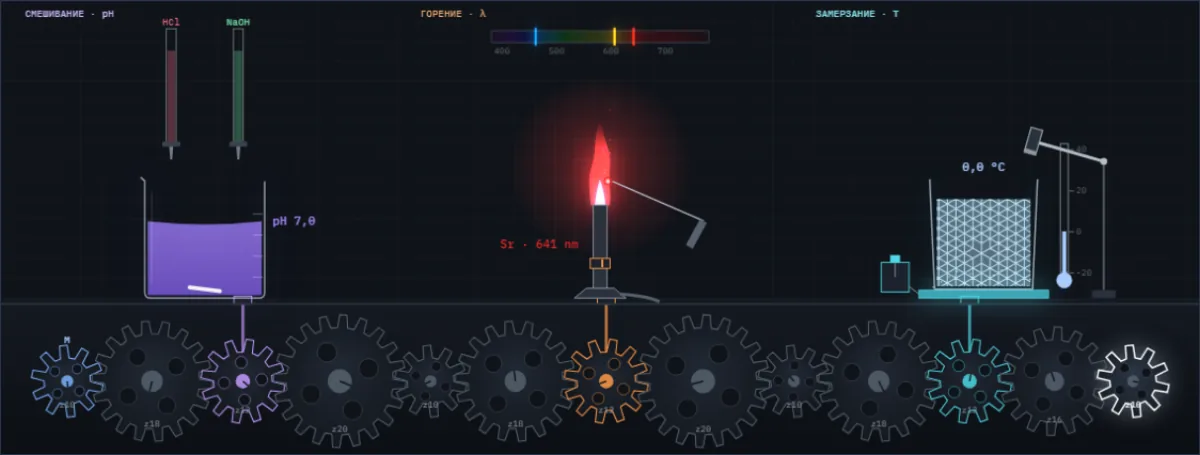
A drop of acid changes the colour of the juice, salt colours the flame, water freezes at a tap. Below them are all Simetrium sheets, by discipline and alphabetically.
Open the index →